Program 08 · Advanced programPlanned
Clinical Imaging & Ultrasound in Aesthetic Medicine
Ultrasound anatomy, image-assisted planning and patient safety
Seeing the anatomy before and during a procedure.
- For
- Physicians with a grounding in aesthetic practice
- Curriculum
- 3 modules · 9 topics
- Format
- To be announced
- Academic direction
- Dr. S. Nouri Rafiee
StatusPlanned. The program is part of the curriculum plan and its content has been outlined; its development, format and dates have not been announced. It is conceived as an advanced program that builds on the core curriculum.
Why imaging belongs in aesthetic training
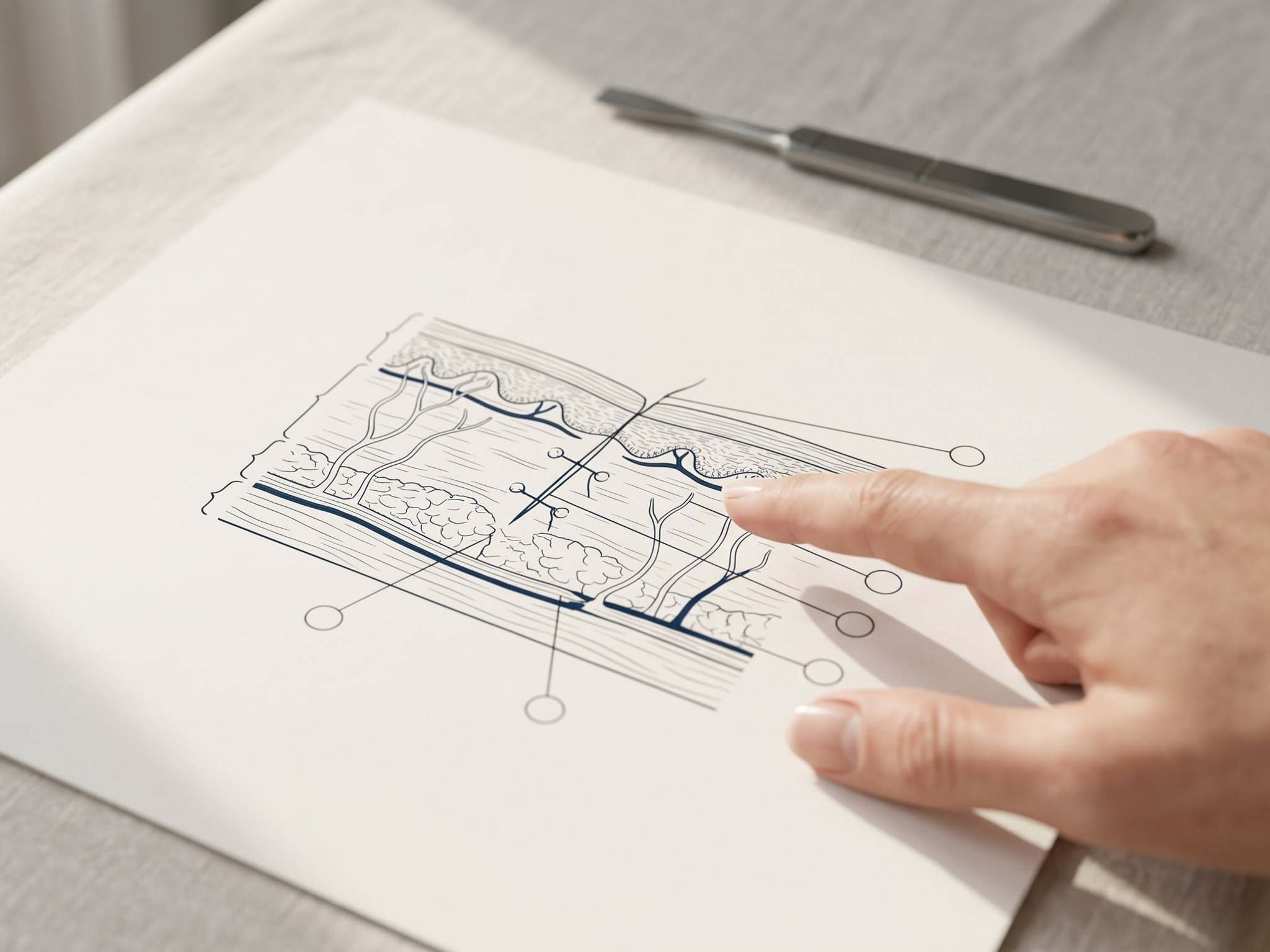
Much of the risk in aesthetic medicine lies in structures that cannot be seen from the surface. The course of a vessel, the depth of a tissue plane or the position of a nerve differs from one patient to the next, and a procedure planned on surface landmarks alone rests on an average that a particular patient may not share. Ultrasound makes part of that anatomy visible before and during a procedure.
It does not make a procedure safe by itself. An image has to be acquired well, read correctly and placed in the context of a clinical decision, and each of those steps can fail. Clinical Imaging & Ultrasound in Aesthetic Medicine is planned to teach imaging in that spirit: as a way of seeing that supports clinical judgment, within the logic of the Rafiee Educational Approach, where assessment and planning come before any intervention.
What the program covers
The program is outlined in three modules. The first is designed to build the reading of the image: facial ultrasound, subcutaneous ultrasound and the vascular anatomy on which the rest of the program depends. The second will turn that reading to the structures at risk, with the identification of vessels and nerves, and to ultrasound-assisted planning, including the findings that should change a plan or stop it. The third will address image-guided procedures, the recognition of complications and the wider safety applications of ultrasound, with escalation and referral taught as part of the same judgment.
The order follows the approach: assess the anatomy, plan with it, and protect the patient before, during and after the procedure.
How it is taught
The program is designed to follow the Academy’s teaching progression. Ultrasound anatomy is to be introduced as foundational knowledge and through demonstration, then practiced on simulation models before any supervised practice, which will take place only where local regulation allows it. Case-based learning is designed to use images without identifiable patient data, so that interpretation and decisions can be discussed openly. Image interpretation matures with repetition, which is why feedback and continued development are part of the design.
Learning will be assessed across knowledge, technical skill, clinical judgment and patient safety, as described in the assessment framework, itself in development. How procedural skills are rehearsed and measured across the curriculum is the subject of a separate planned program, Clinical Simulation, Procedural Skills & Assessment.
How it relates to the other programs
Imaging first appears in Clinical Aesthetic Practice & Patient Safety, where it is introduced as one of the tools of patient safety, alongside consultation, consent and ethics. This program develops imaging and ultrasound in depth, and it assumes the grounding in anatomy and safe practice that the core programs are designed to provide. It also complements Aesthetic Surgical Skills for Physicians: where that program teaches advanced aesthetic technique, this one is concerned with seeing the anatomy that technique has to respect.
The limits are stated plainly. The program does not qualify participants as sonographers, does not replace formal ultrasound training or national requirements, and does not extend anyone’s scope of practice.
Current status
The program is planned: its content has been outlined as part of the curriculum plan, but its development, format and dates have not been announced. It is conceived as an advanced program, to follow the core curriculum. Registering interest is the way to hear when that changes and to tell the Academy how imaging figures in your practice.
An image informs a decision. It does not make a procedure safe on its own.
Who it is for
- Physicians in aesthetic practice who already have a grounding in facial anatomy and safe practice, such as the one the core programs are designed to provide.
- Physicians who use ultrasound in another clinical context and want to understand its applications, and its limits, in aesthetic medicine.
- Physicians interested in imaging as a patient safety tool, as a complement to formal ultrasound training rather than a substitute for it.
Intended outcomes
The program is designed so that participants will be able to:
- Describe how the layers of the face and the subcutaneous tissue appear on ultrasound, and relate them to the vascular anatomy of the regions where aesthetic procedures are performed.
- Recognize on the image the vessels and nerves that matter in a planned procedure, and explain the limits of that recognition.
- Use ultrasound-assisted planning to decide whether, where and how a procedure should go ahead, including the findings that should change the plan or stop it.
- Explain what image guidance can and cannot add during a procedure, and why it does not replace anatomical knowledge or clinical judgment.
- Recognize the imaging features of complications and escalate or refer without delay when a finding is beyond their competence.
- Work within the limits of their training, their license and local regulation, and recognize when a question belongs to a qualified sonographer or radiologist.
Curriculum outline
What the program is designed to cover. The detailed syllabus, format and dates will be published on this page with the first edition.
Where it sits in the Rafiee Educational Approach
- Module 01
Ultrasound anatomy of the face and subcutaneous tissue
Designed to build the reading of the image: how the face and the subcutaneous tissue appear on ultrasound, and the vascular anatomy that every later decision in the program depends on.
- Facial ultrasound
- Subcutaneous ultrasound
- Vascular anatomy
- Module 02
Structures at risk and ultrasound-assisted planning
Turning that reading into a plan: where the vessels and nerves at risk lie in a given patient, and which findings should change a procedure or rule it out.
- Identification of vessels
- Identification of nerves
- Ultrasound-assisted planning
- Module 03
Image guidance, complications and safety
The place of image guidance during a procedure, the recognition of complications and the wider safety applications of ultrasound, with escalation and referral taught as part of the same judgment.
- Image-guided procedures
- Recognition of complications
- Safety applications of ultrasound
How it is taught
Format, dates and venue to be announced. The program is designed to move from foundational knowledge of ultrasound anatomy and demonstration to practice on simulation models, supervised practice where local regulation allows it, and case-based discussion of images, with feedback and assessment throughout.
The teaching progression- 1Foundational knowledge
- 2Demonstration
- 3Simulation
- 4Supervised practice
- 5Case-based learning
- 6Feedback
- 7Assessment
- 8Continued development
Scope and boundaries
What the program does not do matters as much as what it does. These limits are part of the teaching.
- The program does not qualify participants as sonographers. It does not replace formal ultrasound training or the requirements that national regulations set for performing and reporting ultrasound.
- Image guidance does not remove risk. It is taught as an aid to anatomical knowledge and clinical judgment, never as a substitute for them.
- The program does not authorize participants to perform procedures outside their qualifications, their license or their legal scope of practice, which is defined by local regulation. Any procedure discussed is performed only within that scope.
- Suspected injury to vessels, nerves or other deeper structures, and any finding beyond a participant's competence, is taught as recognition and referral, not as a problem to manage independently.
- The program is not currently accredited for CPD or CME credit.
Questions about Clinical Imaging & Ultrasound
Will this program qualify me to perform or report ultrasound?
No. It is designed to teach the applications of ultrasound in aesthetic medicine, not to qualify participants as sonographers. It does not replace formal ultrasound training or the requirements that national regulations set for performing and reporting ultrasound, and nothing in it extends a participant's scope of practice.
Does ultrasound guidance make aesthetic procedures safe?
Not on its own. Ultrasound can show vessels and tissue planes that cannot be seen from the surface, but it does not remove risk. The program is designed to teach imaging as one layer of safety, alongside anatomical knowledge, patient selection, careful technique and the readiness to stop and refer.
How does this program relate to Clinical Aesthetic Practice & Patient Safety?
Program 04 introduces imaging as one of the tools of patient safety, together with consultation, consent and ethics. This program develops imaging and ultrasound in depth. The grounding in safe aesthetic practice that program 04 is designed to provide is the natural preparation for it.
Do I need previous experience with ultrasound?
Entry requirements have not been set. The program is planned as an advanced program that builds on the core curriculum, particularly facial anatomy and patient safety, so it is designed for physicians who already have that grounding rather than as a first introduction to aesthetic practice.
Does the program carry CPD or CME credit?
No. This program is not currently accredited for CPD or CME credit. Any accreditation will be stated on this page only once it has been granted.
All programs
Core programs
The foundation of the curriculum: wound management, procedural basics, aesthetic surgical skills and safe clinical practice.
Wound-to-Scar™
Structured Surgical Wound Management & Scar OptimizationFlagship · In developmentFoundations of Clinical & Minimally Invasive Procedural Medicine
Fundamental clinical and procedural skillsIn developmentAesthetic Surgical Skills for Physicians
Aesthetic analysis, local flaps, plasties and fine techniqueIn developmentClinical Aesthetic Practice & Patient Safety
Safe clinical practice and decision-making in aesthetic medicineIn development
Advanced & specialized programs
Programs that build on the core: simulation and assessment, consultation and practice, and clinical imaging.
Clinical Simulation, Procedural Skills & Assessment
How procedural skills are learned, rehearsed and assessedPlannedAesthetic Consultation, Ethics & Practice Development
Communication, consent, ethical conduct and the organization of a practicePlannedClinical Imaging & Ultrasound in Aesthetic Medicine
Ultrasound anatomy, image-assisted planning and patient safetyPlanned
Research & educational development
A line of inquiry into the biology of healing and scarring. It studies the science behind the programs and is not taught.